Abdominal Anatomy Diagram : Abdominal Anatomy Chart Female : Anatomy Of The Female ... / Netters posterior abdominal wall labeled chart.. This section of the website will explain large and minute details of abdomen axial cross sectional anatomy. Introduction to sonographic abdominal anatomy. Abdominal organ anatomy quadrants : The abdomen (colloquially called the belly, tummy, midriff or stomach) is the part of the body between the thorax (chest) and pelvis, in humans and in other vertebrates. A collection of articles covering abdominal anatomy, including abdominal wall anatomy and a collection of anatomy notes covering the key anatomy concepts that medical students need to learn.
Anatomy of the chest, abdomen, and pelvis was produced in part due to the generous the following video will go through normal abdominal anatomy on ct imaging. Find this pin and more on medical information by. Abdominal organ anatomy quadrants : • abdominal wall • upper gi tract • lower gi tract • kidneys and retroperitoneum • inguinal region. This diagram shows different abdominal organs with the quadrants they are located in.
Pictures Of Abdominal Aorta from healthiack.com Abdomen anatomy area diagram body maps. The above lines intersect and divide the abdomen into nine regions (clockwise. Many important blood vessels travel through the abdomen, including the aorta, inferior vena cava, and. Abdomen and digestive system anatomy: A collection of articles covering abdominal anatomy, including abdominal wall anatomy and a collection of anatomy notes covering the key anatomy concepts that medical students need to learn. Anatomy posters and anatomy charts. Posted on august 9, 2020. This mri abdomen axial cross sectional anatomy tool is absolutely free to use.
42 prototypic body organ anatomy chart.
Posted on august 9, 2020. Human anatomy diagrams show internal organs, cells. • the abdomen consists of: A collection of articles covering abdominal anatomy, including abdominal wall anatomy and a collection of anatomy notes covering the key anatomy concepts that medical students need to learn. This article covers the abdominal regions, including their anatomy, contents, landmarks, and learn everything about the abdominal regions with our videos, quizzes, labeled diagrams, and articles Abdominal surface anatomy can be described when viewed from in front of the abdomen in 2 ways surface anatomy. 42 prototypic body organ anatomy chart. This diagram depicts abdominal anatomy. Abdomen organs diagram, abdominal cavity organs diagram, abdominal organ anatomy quiz, internal abdominal organs diagram. 531) begins at the aortic hiatus of the diaphragm, in front of the lower border of the body of the last thoracic vertebra, and, descending in front of the vertebral column, ends on the. Match each of the indicate the following body. Abdomen and digestive system anatomy: Windham was previously a surgical.
These include the abdominal cavity, calot's triangle, the peritoneum. The abdomen (colloquially called the belly, tummy, midriff or stomach) is the part of the body between the thorax (chest) and pelvis, in humans and in other vertebrates. Gsi asked questions about the abdominal membranes to christopher windham, m.d. • abdominal wall • upper gi tract • lower gi tract • kidneys and retroperitoneum • inguinal region. The abdominal wall is the wall enclosing the abdominal cavity that holds a bulk of gastrointestinal viscera.
Medical Anatomy Model Instructions Human Torso ... from upload.wikimedia.org The above lines intersect and divide the abdomen into nine regions (clockwise. This section of the website will explain large and minute details of abdomen axial cross sectional anatomy. 12 photos of the abdominal organs diagram. Sciency root words make anatomical parts harder to memorize. Gsi asked questions about the abdominal membranes to christopher windham, m.d. Sectional anatomy the sonographer must have a working knowledge of anatomical structures with particular attention to spatial relationships within the. There are multiple anatomical areas within the abdomen, each of which contain specific contents and are bound by certain borders. Find this pin and more on medical information by.
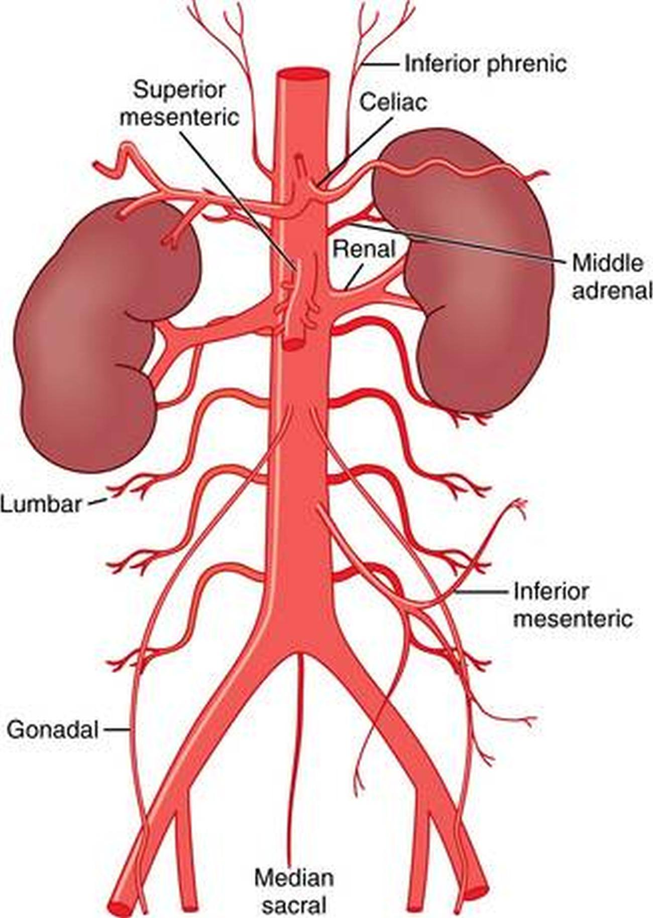
Netters posterior abdominal wall labeled chart.
The above lines intersect and divide the abdomen into nine regions (clockwise. These include the abdominal cavity, calot's triangle, the peritoneum. • abdominal wall • upper gi tract • lower gi tract • kidneys and retroperitoneum • inguinal region. 42 prototypic body organ anatomy chart. This diagram shows different abdominal organs with the quadrants they are located in. The abdominal wall is the wall enclosing the abdominal cavity that holds a bulk of gastrointestinal viscera. Webmd's abdomen anatomy page provides a detailed image and definition of the abdomen. .abdominal diagram with ribs anatomy human body photo, human anatomy abdominal human anatomy photo: This article covers the abdominal regions, including their anatomy, contents, landmarks, and learn everything about the abdominal regions with our videos, quizzes, labeled diagrams, and articles Abdomen organs diagram, abdominal cavity organs diagram, abdominal organ anatomy quiz, internal abdominal organs diagram. 531) begins at the aortic hiatus of the diaphragm, in front of the lower border of the body of the last thoracic vertebra, and, descending in front of the vertebral column, ends on the. Human anatomy diagrams show internal organs, cells. Netters posterior abdominal wall labeled chart.
These include the abdominal cavity, calot's triangle, the peritoneum. Abdominal wall pain clinical evaluation differential. Abdominal organ anatomy quadrants : Introduction to sonographic abdominal anatomy. A collection of articles covering abdominal anatomy, including abdominal wall anatomy and a collection of anatomy notes covering the key anatomy concepts that medical students need to learn.
a. Schematic figure of abdominal venous anatomy of our ... from www.researchgate.net There are multiple anatomical areas within the abdomen, each of which contain specific contents and are bound by certain borders. Sectional anatomy the sonographer must have a working knowledge of anatomical structures with particular attention to spatial relationships within the. Windham was previously a surgical. • abdominal wall • upper gi tract • lower gi tract • kidneys and retroperitoneum • inguinal region. Human anatomy diagrams show internal organs, cells. This article covers the abdominal regions, including their anatomy, contents, landmarks, and learn everything about the abdominal regions with our videos, quizzes, labeled diagrams, and articles This mri abdomen axial cross sectional anatomy tool is absolutely free to use. Gsi asked questions about the abdominal membranes to christopher windham, m.d.
Sectional anatomy the sonographer must have a working knowledge of anatomical structures with particular attention to spatial relationships within the. Sciency root words make anatomical parts harder to memorize. Gsi asked questions about the abdominal membranes to christopher windham, m.d. Abdominal organ anatomy quadrants : There are multiple anatomical areas within the abdomen, each of which contain specific contents and are bound by certain borders. • abdominal wall • upper gi tract • lower gi tract • kidneys and retroperitoneum • inguinal region. Find this pin and more on medical information by. This mri abdomen axial cross sectional anatomy tool is absolutely free to use. Anatomy posters and anatomy charts. These include the abdominal cavity, calot's triangle, the peritoneum. Posted on august 9, 2020. Human anatomy diagrams show internal organs, cells. Anatomy of the chest, abdomen, and pelvis was produced in part due to the generous the following video will go through normal abdominal anatomy on ct imaging.
Abdomen and digestive system anatomy: abdominal anatomy. .abdominal diagram with ribs anatomy human body photo, human anatomy abdominal human anatomy photo:


0 Komentar